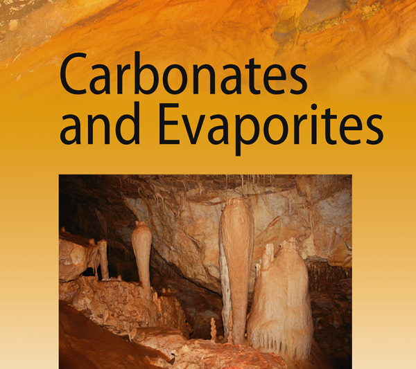
18th March 2016

Badley Ashton Presentation at the 2016 Janet Watson Meeting


Badley Ashton's Research and Business Development Manager, Adrian Neal will be presenting at the 2016 Janet Watson Meeting - The Future of Hydrocarbon Exploration. His presentation - "Detailed reservoir characterization using 3D CT scans, borehole imaging and core" is based on ongoing work by our Houston based Deepwater team in conjunction with our clients at BP and highlights the advantages that can be gained from an analytical approach that combines the use of 3D CT scans with more traditional techniques, you can read the abstract below. Badley Ashton are also proud to sponsor this event along with Dong Energy and Shell. The event will be held on the 27th and 28th of April at Burlington House in London.

The presentation is scheduled for 14:40 on Thursday the 28th of April, but Adrian will be in attendance for both days. He will be very happy to discuss how Badley Ashton can help you in all aspects of reservoir geology, so please come and say hello. We look forward to seeing you there.


Detailed reservoir characterization using 3D CT scans, borehole imaging (BHI) and core

Computer Axial Tomography (CAT or CT scanning), is an X-ray based technique first developed in the late 1960s for medical '3D' imaging purposes.  However, it has increasingly found applications in the earth sciences due to its ability to provide both quantitiative and qualititaive data regarding the internal structure of rocks and sediments, as determined by contrasts in density and atomic composition.  The integration of 3D CT Scans from core with more traditional core description techniques, and other technologies such as borehole imaging (BHI), offers a powerful way of characterising sedimentological and structural features in a reservoir.  With borehole images it is possible to identify and orient geologic features over relatively long well sections (100s - 1000s of feet). However, BHI resolution is still very coarse compared to what can be observed in a core, which offer a very detailed picture of the reservoir rock.  Despite this, subtle sedimentary and structural features can be difficult to see with the naked eye, especially if the core is oil stained. In these cases the use of 3D CT scans offers a very high resolution image of the structures in the rock, which can be used to enhance the core description.  The study we present here shows a detailed interpretation combining 3D CT scans, BHI and core from a deepwater turbidite reservoir.  Firstly, the 3D CT scans allowed more detailed recognition of sedimentary facies; commonly sandstones that seemed structureless in the slabbed core were shown to be space-laminated or dewatered in the CT scans.  Secondly, by using bedding features common to both the BHI and core data sets, it was possible to orient the 3D CT scans and demonstrate that interpreted fractures in the BHI corresponded to deformation band sets in the core.  Using the CT scans it was possible to identify more deformation bands than with the BHI.  However, most of the fractures identified with the BHI matched both in orientation and location those interpreted in the CT scans. Thus by combining CT scans and BHI it is possible to orient sedimentological and structural features to provide a more complete picture of the reservoir at a particular well location, and provide calibration for wells where only BHI are available.


LinkedIn Button Email to Friend Button